

 Türkçe
Endocrine surgery is a specialized field that covers the surgical treatment of diseases of hormone-producing glands such as the thyroid, parathyroid, adrenal, and pancreas. This branch of surgery includes procedures aimed at removing masses and tumors that disrupt hormonal balance.
Thyroid surgery is the most commonly performed part of endocrine surgery and is typically carried out for the treatment of thyroid nodules, goiter, or thyroid cancer. The aim of the operation is to halt disease progression while preserving hormonal balance and reducing the risk of complications.
Parathyroid surgery is performed to treat hyperparathyroidism, which develops when the parathyroid glands—responsible for regulating calcium levels in the blood—work excessively. This operation provides positive effects on bone health and kidney function.
Adrenal and pancreatic surgeries are critically important for treating rare but serious tumors that can cause significant hormonal disorders. These procedures may be performed using minimally invasive techniques, helping to speed up the patient’s recovery process.
| Key Information | Details |
| Definition of Endocrine Surgery | A medical specialty that involves the surgical treatment of diseases developing in hormone-producing glands (thyroid, parathyroid, adrenal glands, and certain pancreatic/liver foci). |
| Conditions That May Require Surgery | Thyroid nodules, thyroid cancers, hyperthyroidism, parathyroid adenomas, primary hyperparathyroidism, adrenal tumors, functional pancreatic tumors such as insulinoma and gastrinoma. |
| Common Endocrine Operations | Thyroidectomy (removal of the thyroid gland), parathyroidectomy, adrenalectomy (removal of the adrenal gland), resections of endocrine pancreatic tumors. |
| Thyroid Surgery | Removal of part or all of the thyroid gland due to nodules, goiter, hyperthyroidism, or cancer. |
| Parathyroid Surgery | Surgical removal of an adenoma that secretes excess hormone (usually the cause of primary hyperparathyroidism). |
| Adrenal (Suprarenal) Surgery | Removal of hormone-producing or enlarging tumors; helps resolve problems such as high blood pressure and weight gain caused by hormone excess. |
| Endocrine Pancreatic Surgery | Removal of hormone-secreting neuroendocrine tumors such as insulinoma, gastrinoma, and glucagonoma. |
| Diagnostic Methods | Hormone tests, biochemical analyses, ultrasonography, CT, MRI, scintigraphy, PET, fine-needle aspiration biopsy, genetic analyses. |
| Preoperative Assessment | A multidisciplinary assessment is performed together with an endocrinology specialist; the patient’s hormonal balance is optimized to make surgery safe. |
| Surgical Techniques | Open surgery, laparoscopic and robotic techniques; the chosen method depends on the location, size, and spread of the disease. |
| Postoperative Follow-up | Regular monitoring of hormone levels, wound healing, vocal cord function, and calcium balance is required. |
| Possible Complications | Vocal cord nerve injury (especially in thyroid and parathyroid surgeries), bleeding, infection, temporary or permanent hypocalcemia. |
| Long-Term Follow-up | Ongoing control of the disease and monitoring for recurrence through hormone therapy, imaging, and laboratory tests. |
| Multidisciplinary Approach | Endocrine surgery is carried out in close collaboration with endocrinology, nuclear medicine, radiology, pathology, and medical oncology. |


You can get information about Endocrine Surgery prices, the process, and the hospital where Endocrine Surgery is performed by contacting Assoc. Prof. Dr. Aydıncan, one of the doctors performing Endocrine Surgery in Ankara.

Assoc. Prof. Dr. Aydıncan Akdur
General Surgery Specialist
General Surgery Specialist
I graduated from Cumhuriyet University Faculty of Medicine in 2002. In 2009, I completed my General Surgery specialization at Başkent University. After working at Elmadağ State Hospital, I began working at Başkent University; I became Assistant Professor in 2014 and Associate Professor in 2021. Between 2015 and 2022, I served as the Responsible Physician of the Kidney Transplant Center at Başkent University Konya Hospital. I have participated in more than 1,000 kidney transplants, nearly 300 liver transplants, and close to 2,500 surgeries.
Between 2022 and 2025, I provided services at Bayındır Healthcare Group.
Since 2023, I have been serving as the Head of the Department of General Surgery at Ankara Medipol University.
I am actively accepting patients at Ankara Private 100th Year Hospital and my private clinic.
About Me ContactWhat Is Endocrine Surgery?
Endocrine surgery is a surgical specialty that deals with the operative treatment of diseases affecting hormone-producing glands (the thyroid, parathyroid, adrenal glands, and certain parts of the pancreas). Surgeries in this field most commonly include thyroid nodules, thyroid and parathyroid diseases, adrenal gland tumors, and endocrine pancreatic tumors. Endocrine surgery is a field that requires high precision in terms of hormonal balance and oncologic outcomes and is carried out through multidisciplinary collaboration.
In Which Situations Is Thyroid Surgery Necessary?
Located at the front of the neck and shaped like a butterfly, the thyroid gland is the main control center of our metabolism. There are important situations in which operating on this small organ becomes necessary. These situations generally arise when there is a cancer risk, when hormonal balance is disrupted, or when the gland causes physical discomfort. The main conditions that lead to a decision for thyroid surgery include:
- Thyroid Cancer
- Goiter Causing Compression
- Toxic Goiter (Hyperthyroidism)
- Suspicious Nodules
Each of these headings helps us better understand why surgical intervention may be required. Thyroid cancer—or a strong suspicion of cancer—is the clearest and most important reason for surgery. The goal is to achieve an oncologic victory by completely removing cancerous tissue and, if it has spread, the nearby at-risk lymph nodes.
Goiter refers to enlargement of the thyroid gland. Sometimes this enlargement becomes so advanced that it begins to compress the windpipe or esophagus. This can cause difficulty swallowing, shortness of breath, voice changes, or a persistent pressure sensation in the neck. In addition, goiters that create a visible swelling from the outside (causing cosmetic concerns) or that grow downward into the chest and may lead to more serious problems are also removed surgically.
Toxic goiter (hyperthyroidism) is a condition in which the thyroid gland produces excessive hormones uncontrollably. This turns the body’s metabolism into an engine running at full speed and causes symptoms such as palpitations, excessive sweating, weight loss, irritability, and tremors in the hands. When medication is ineffective, side effects cannot be tolerated, or a definitive solution is desired, surgical removal of this overactive hormone-producing tissue is the most effective treatment.
Finally, suspicious nodules are also an important reason for surgery. Sometimes a fine-needle aspiration biopsy does not yield a definitive answer and is reported as “indeterminate” or “suspicious.” In such cases, the only way to establish a definitive diagnosis—whether the nodule is benign or malignant—is to surgically remove the thyroid lobe containing the nodule and send it for pathological examination. This can be considered a “diagnostic surgery.”
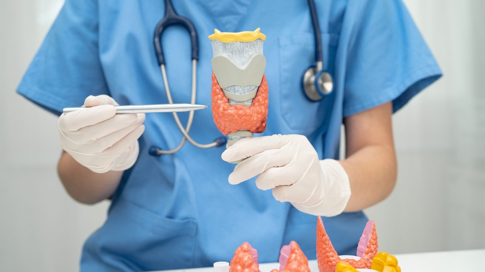
What Evaluations Are Performed Before Deciding on Thyroid Surgery?
Deciding whether a thyroid nodule should be operated on is much like a detective piecing together clues. Every piece of data—from the patient’s history and blood tests to imaging findings and biopsy results—is carefully combined to reach the correct diagnosis and treatment plan. This process ensures the most appropriate step is taken for the patient’s health.
One of the first and most important steps is measuring the level of Thyroid Stimulating Hormone (TSH) in your blood. You can think of TSH as a boss in the brain giving the thyroid gland the command to “work” or “stop.” If the TSH level is low, it indicates the thyroid is overworking on its own. These “hot nodules” have an almost zero risk of cancer. However, if TSH is normal or high, it suggests the nodule is not producing hormones (“cold nodule”) and the cancer risk may be higher, prompting the next step.
That next step is high-resolution thyroid ultrasonography. Ultrasound creates a detailed map of the thyroid gland and the nodules within it. On this map, we look for suspicious signs that increase the likelihood of malignancy. Some suspicious ultrasound features that may raise the risk of cancer include:
- Dark appearance (Hypoechoic)
- Irregular, spiculated margins
- Tiny calcifications (Microcalcifications)
- Taller-than-wide shape
- A firm, solid structure
Ultrasound also evaluates neck lymph nodes, providing valuable clues about whether cancer has spread.
If suspicious findings are detected on ultrasound, the gold standard Fine-Needle Aspiration Biopsy (FNAB) is performed. In this procedure, a very thin needle is inserted into the nodule under ultrasound guidance to collect a small sample of cells. A pathologist examines these samples under a microscope. The procedure is usually quick and painless. The biopsy result provides highly accurate information about the nature of the nodule and largely clarifies the question of “surgery or follow-up?”
Which Surgical Techniques Are Used for Thyroid Surgery?
Thyroid surgery has undergone major changes in recent years with advances in technology and surgical principles. Instead of a single standard method, there are now different approaches that can be selected based on the patient’s condition, the type of disease, and even cosmetic expectations.
Conventional Open Thyroidectomy is still the most commonly used and remains the “gold standard” that can be safely applied to all types of thyroid disease. In this technique, the thyroid gland is reached through a horizontal incision made along a natural skin crease in the lower neck. Its greatest advantage is that it provides the surgeon with a wide field of view and working space, making it easier to safely identify and protect the most critical structures—such as the nerves controlling the vocal cords and the tiny parathyroid glands responsible for calcium balance. This method is indispensable especially for large goiters, thyroid cancers, and cases requiring removal of lymph nodes in the neck.
Today, however, cosmetic concerns are also considered part of treatment success. In response, techniques that leave less visible scarring—or none at all—have been developed. Minimally Invasive Video-Assisted Thyroidectomy (MIVAT) uses a much smaller incision (about 1.5–3 cm) and is performed with the help of a camera. It offers better cosmetic results but is only suitable for selected patients with small nodules and smaller thyroid glands.
One of the most recent and exciting developments in scarless thyroid surgery is Transoral Endoscopic Thyroidectomy Vestibular Approach (TOETVA), commonly referred to as “scarless thyroid surgery through the mouth.” With this technique, no incision is made on the skin. The entire procedure is carried out through three small openings made inside the lower lip, in an area that no one can see. The thyroid gland is removed using a camera and long, thin instruments. The cosmetic outcome is excellent. However, this advanced technique is not suitable for everyone and is only performed in carefully selected patients with small benign nodules or early-stage small cancers. The choice of technique is a shared decision based on the condition of the disease and the surgeon’s experience.
Get in Touch with Us.
Click the button to contact us and request an online appointment. Our team will get in touch with you as soon as possible.
Why and When Is Parathyroid Surgery Necessary?
Hidden just behind the thyroid gland, the parathyroid glands—usually four lentil-sized glands—act as the body’s calcium thermostats. Their job is to keep blood calcium levels in a precise balance by secreting Parathyroid Hormone (PTH). Sometimes, one or more of these glands becomes overactive due to a benign tumor (adenoma) and starts producing excessive PTH. This condition is called “Primary Hyperparathyroidism” and can completely disrupt the body’s calcium balance.
Persistently high calcium levels in the blood may progress silently at first, but over time they can lead to serious health problems. Unwanted effects appear in different parts of the body. Some common problems caused by hyperparathyroidism include:
- Kidney stones
- Bone loss (Osteoporosis) and bone pain
- Chronic weakness and fatigue
- Memory problems and difficulty concentrating
- Digestive complaints and constipation
- Depression and mood changes
There is no definitive, permanent medical (drug) treatment for this condition. The only certain and lasting way to stop this “calcium storm” in the body is to surgically remove the diseased parathyroid gland(s) producing excess hormone. Surgery often results in a dramatic improvement in quality of life and prevents serious health problems associated with prolonged high calcium levels.
How Is the Diseased Parathyroid Gland Located Before Surgery?
The key to a successful parathyroid operation is accurately identifying the diseased gland before entering the operating room. This process is like a “treasure hunt,” and the surgeon uses modern maps to define the target in advance. This makes it possible to plan a small, focused operation instead of exploring the entire neck.
Two main “maps” are typically used in this treasure hunt. The first is neck ultrasonography, which can visualize an enlarged, abnormal parathyroid adenoma. The second is a nuclear medicine test called Sestamibi Scintigraphy. In this test, a special radioactive substance is injected into a vein. This substance is taken up mainly by the overactive parathyroid gland. On the scan images, the diseased gland lights up like a bulb, revealing its location.
When these two tests match—meaning both ultrasound and scintigraphy point to the same spot—the gland’s location can be identified with over 95% accuracy. This is usually sufficient to plan minimally invasive surgery.
Sometimes, however, these initial maps may not be conclusive or may conflict. In difficult cases—especially in patients who have had prior surgery but still have disease, or when the gland is suspected to be in an unusual (ectopic) location—more advanced mapping is needed. 4D Computed Tomography (4D-CT) is a highly sensitive imaging method that shows the adenoma’s blood flow characteristics in detail. With this advanced technology, even the best-hidden parathyroid adenomas can be detected.
Which Methods Are Used During Parathyroid Surgery?
Parathyroid surgery is an elegant operation shaped by the clarity of the “treasure maps” we have. Today, the standard approach for most patients is Minimally Invasive Parathyroidectomy (MIP). In this technique, a small incision of about 2.5–3.5 cm is made directly over the area where the diseased gland is located. Through this small window, the surgeon gently removes the previously localized diseased gland (adenoma). This targeted approach means less tissue trauma, less pain, faster recovery, and excellent cosmetic outcomes.
A very important technological aid that maximizes the success and safety of these minimally invasive surgeries is the Intraoperative Rapid PTH Test (ioPTH). PTH has a very short half-life in the blood—only a few minutes. By taking advantage of this, a blood sample is drawn again about 10 minutes after the diseased gland is removed, and the PTH level is measured. If the PTH level drops by more than 50% compared to the baseline, it confirms that the surgery has been successful and that no additional overactive gland is present. This test gives the surgeon a real-time “success confirmation.” If the PTH level does not drop sufficiently, it suggests a second adenoma or multigland disease, and the surgeon can expand the operation in the same session to evaluate the other glands. This method serves as a safety valve that helps guarantee the high success rates of minimally invasive parathyroid surgery.
In rare cases where imaging is inconclusive or multigland disease is suspected, the traditional four-gland exploration—finding and evaluating all four parathyroid glands—may still be performed.
Get in Touch with Us.
You can request an online appointment by filling out the form. Our team will contact you as soon as possible.
Which Diseases Require Adrenal Gland Surgery?
Sitting like caps on top of each kidney, the adrenal glands produce powerful hormones that regulate vital functions such as stress response, blood pressure, blood sugar, and mineral balance. Tumors that develop in these glands either disrupt the body’s chemistry by producing excess hormones or raise suspicion for cancer based on their size and features. Adrenal gland surgery (adrenalectomy) is generally considered in these two scenarios. There are several types of functional tumors that cause hormone overproduction. The most common include:
- Conn Syndrome (Excess Aldosterone)
- Cushing Syndrome (Excess Cortisol)
- Pheochromocytoma (Excess Adrenaline)
Conn syndrome is characterized by excessive aldosterone production and often causes treatment-resistant high blood pressure. Cushing syndrome results from excess cortisol and can lead to weight gain, high blood sugar, and easy bruising. Pheochromocytoma secretes excessive adrenaline and noradrenaline, causing attacks of severe headache, palpitations, sweating, and potentially life-threatening hypertensive crises. All of these functional tumors must be surgically removed to calm the hormonal storm they create.
Another important reason for surgery is suspicion of cancer. The size of an adrenal mass provides an important clue about malignant potential. In general, the risk of cancer increases in masses larger than 4 cm, and becomes quite significant in masses exceeding 6 cm. In selected patients, surgical removal may also be an option when another cancer has spread (metastasized) to the adrenal gland.
What Tests Are Done for an Incidentally Discovered Adrenal Mass (Incidentaloma)?
Today, it is fairly common to detect an adrenal mass incidentally on a CT or MRI performed for another reason. These are called “adrenal incidentalomas” and they initiate a careful evaluation process for both patient and physician. There is no need to panic, because the vast majority of these masses are benign and harmless. The key is to distinguish harmless lesions from those requiring surgery. For this reason, we systematically seek answers to two main questions:
Is this mass producing hormones? Blood and urine tests are mandatory to answer this question. Every patient is screened for Cushing syndrome, Conn syndrome, and pheochromocytoma. Among these, excluding pheochromocytoma is especially critical. Because any intervention performed without recognizing this tumor (such as a biopsy) can trigger a fatal hypertensive crisis. Therefore, the rule is: “first hormone tests, then everything else.”
Is there a risk that this mass is malignant? The answer largely depends on imaging characteristics, especially the appearance on a non-contrast CT scan. Most benign adrenal lesions contain a high amount of fat. On CT, this fatty tissue appears as low density (10 Hounsfield Units or less). This finding is strong evidence that the mass is benign. If the density is higher or the size is greater than 4 cm, the risk of cancer increases and surgery is often recommended.
A roadmap is drawn based on these answers. If the mass is hormone-producing or suspicious for cancer, it is removed surgically. If it does not produce hormones and shows clearly benign features, periodic follow-up is usually sufficient.
Which Surgical Methods Are Used for Adrenal Surgery?
The choice of method in adrenal surgery is primarily determined by whether the tumor is benign or malignant. This decision is vital for both oncologic success and patient safety.
Open Adrenalectomy is the traditional approach performed through a large abdominal incision. Today, it is reserved for specific situations—most importantly for known or suspected adrenocortical carcinoma (ACC), a type of adrenal cancer. A fundamental principle in cancer surgery is removing the tumor in one piece with surrounding tissue, without rupturing the capsule (en-bloc resection). Capsule rupture can spill cancer cells into the abdomen and lead to disease spread. Open surgery provides the wide exposure and control needed to follow this principle safely. It is also preferred for very large tumors (often larger than 10–12 cm).
For most benign adrenal tumors, the “gold standard” treatment is Laparoscopic (Minimally Invasive) Adrenalectomy. In this method, a camera and fine surgical instruments are inserted through 3–4 small (about 1 cm) incisions made in the abdomen or back. The surgeon performs the operation while viewing a magnified image on a screen. The advantages of this minimally invasive approach are clear: much less postoperative pain, shorter hospital stay (usually 1–2 days), faster return to daily life and work, and better cosmetic outcomes.
There are two main laparoscopic approaches: Transabdominal (through the abdomen) and Posterior Retroperitoneoscopic (through the back). The posterior approach reaches the adrenal gland directly without entering the abdominal cavity and can offer major advantages, especially in patients who have had prior abdominal surgery or in those with obesity. However, because it is technically more demanding, it is typically performed by surgeons in high-volume centers with special expertise.
When Is Surgical Treatment Necessary for Pancreatic Neuroendocrine Tumors?
Neuroendocrine tumors arising from the hormone-producing cells of the pancreas (pNETs) are a rare and unique group of tumors that can behave very differently. Some grow very slowly and cause no symptoms for years, while others can be more aggressive. Therefore, the treatment decision is individualized—like tailoring a suit—based on many factors such as whether the tumor produces hormones (functional vs non-functional), its size, and its location within the pancreas.
Functional pNETs cause clinical symptoms due to the hormones they produce. For example, an insulinoma that produces excess insulin leads to recurrent low blood sugar attacks, while a gastrinoma that produces excess gastrin can cause severe peptic ulcers. These tumors should be surgically removed regardless of size to treat the hormonal syndrome they create.
Non-functional pNETs do not produce hormones and are often discovered incidentally on imaging performed for other reasons. In such cases, the decision for surgery largely depends on the tumor’s size and therefore its malignant potential. The general approach is: tumors larger than 2 cm have a higher cancer risk and are recommended for surgical removal. Tumors smaller than 1 cm are often slow-growing benign lesions and can be managed with “active surveillance” using periodic imaging. Tumors between 1 and 2 cm represent a “gray zone.” In these cases, the decision is individualized by considering factors such as the tumor’s location, the patient’s overall health, and patient preferences.
How Does the Recovery Process Work After Endocrine Surgery?
Careful follow-up and proper postoperative care are as important as a successful operation. This process is designed to detect and manage potential problems early.
The first 24–48 hours after surgery require the closest monitoring. After thyroid and parathyroid (neck) surgery, one of the key concerns is bleeding (hematoma) in the surgical area. Although rare, accumulating blood can compress the windpipe. Therefore, patients are closely observed for symptoms such as sudden neck swelling, shortness of breath, or a pressure sensation in the neck. After adrenal surgery, especially when a hormone-producing tumor has been removed, fluctuations in blood pressure may occur and close blood pressure monitoring is needed.
After discharge, there are also warning signs that patients should watch for. If any of the following occur, you should contact your surgeon immediately. These “red flag” symptoms include:
- Sudden and progressive swelling of the neck
- Difficulty breathing or wheezing
- Severe numbness, tingling, or cramping in the hands, feet, or around the lips
- Fever above 38.5°C (101.5°F)
- Redness, increased warmth, or purulent discharge from the surgical wound
These symptoms may indicate a problem that requires urgent medical attention.
Frequently Asked Questions
Which conditions are covered by endocrine surgery?
Endocrine surgery covers diseases of hormone-producing organs such as the thyroid, parathyroid, adrenal (suprarenal) glands, and the pancreas. Nodules, tumors, cysts, and conditions that cause hormonal imbalances fall within this scope.
When is endocrine surgery necessary?
Endocrine surgery may be required in conditions such as a risk of malignancy in thyroid nodules, overactivity of the parathyroid glands, or adrenal gland tumors. Surgery is usually performed when medical treatment is insufficient.
What is the recovery process like after thyroid surgery?
After thyroid surgery, patients usually stay in the hospital for 1–2 days. The average recovery time is about 1–2 weeks. Hoarseness or mild difficulty swallowing may be temporary. Thyroid hormone levels should be monitored regularly.
Which diagnostic methods are used in endocrine surgery?
Methods such as ultrasound, biopsy, blood tests, hormone analyses, and scintigraphy are used. These tests help detect structural changes in the glands and disorders of hormone production.
How is hormonal balance maintained after endocrine surgery?
After surgery, some hormone deficiencies may occur. In such cases, hormone replacement therapy is administered under medical supervision. Hormone levels are kept under control through regular blood tests.
What should be considered after parathyroid surgery?
After parathyroid surgery, calcium levels are closely monitored. Muscle cramps in the hands and face can occur due to low calcium. In such cases, supplementation is started and calcium-rich foods are recommended.
How are adrenal gland tumors treated in endocrine surgery?
In adrenal tumors, the surgical approach is generally performed laparoscopically. This method involves smaller incisions and a faster recovery period. Hormonal balance is closely monitored after surgery.
What are the risks and complications in endocrine surgery?
Hoarseness, bleeding, infection, or hormonal imbalances are possible complications in endocrine surgery. When performed by experienced surgeons, these risks are quite low.
Why does hoarseness occur after endocrine surgery?
In thyroid or parathyroid surgeries, temporary hoarseness may occur if nerves near the vocal cords are affected. In most cases, the voice returns to normal within a few weeks; rarely, it can be permanent.
How often should follow-up visits be scheduled after endocrine surgery?
The first follow-up is usually performed within 1–2 weeks after surgery. After that, follow-up every 3–6 months may be recommended depending on hormone levels and overall health. These visits are important for maintaining long-term balance.